


Images
 Western blot - Anti-beta III Tubulin antibody - Neuronal Marker (ab18207)
Lane 1: Wild-type HAP1 whole cell lysate (20 µg)Lane 2:Beta III Tubulin knockout HAP1 whole cell lysate (20 µg)
Lanes 1 - 2: Merged signal (red and green). Green - ab18207 observed at 55 kDa. Red - loading control, ab9484, observed at 37 kDa.
ab18207 was shown to recognize beta III Tubulin in wild-type HAP1 cells as signal was lost in beta III Tubulin knockout cells. An additional cross-reactive band at 50 kDa was observed in wild-type and knockout cells. Due to the immunogen’s homology with TUB (Tubby protein homolog, Uniprot: P50607), this lower band could correspond to the TUB protein. Please note that cross-reactivity with this protein has not been confirmed experimentally.
Wild-type and beta III Tubulin knockout samples were subjected to SDS-PAGE. Ab18207 and ab9484 (Mouse anti-GAPDH loading control) were incubated overnight at 4°C at 1 μg/ml and 1/20000 dilution respectively. Blots were developed with Goat anti-Rabbit IgG H&L (IRDye® 800CW) preabsorbed ab216773 and Goat anti-Mouse IgG H&L (IRDye® 680RD) preabsorbed ab216776 secondary antibodies at 1/20000 dilution for 1 hour at room temperature
 Immunocytochemistry/ Immunofluorescence - Anti-beta III Tubulin antibody - Neuronal Marker (ab18207)
ab18207 staining beta III Tubulin in SK-N-SH (Human neuroblastoma cell line) cells. The cells were fixed with 100% methanol (5min) and then blocked in 1% BSA/10% normal goat serum/0.3M glycine in 0.1%PBS-Tween for 1h. The cells were then incubated with ab18207 at 1μg/ml and ab7291 at 1µg/ml overnight at +4°C, followed by a further incubation at room temperature for 1h with a Goat Anti-Rabbit IgG H&L (Alexa Fluor® 488) preadsorbed (ab150081) secondary antibodyat 2 μg/ml (shown in green) and Goat Anti-Mouse IgG H&L (Alexa Fluor® 594) (ab150120) secondary antibodyat 2 μg/ml (shown in pseudo color red). Nuclear DNA was labelled in blue with DAPI.Negative controls: 1– Rabbit primary and anti-mouse secondary antibody; 2 – Mouse primary antibody and anti-rabbit secondary antibody. Controls 1 and 2 indicate that there is no unspecific reaction between primary and secondary antibodies used.
 Immunohistochemistry (Formalin/PFA-fixed paraffin-embedded sections) - Anti-beta III Tubulin antibody - Neuronal Marker (ab18207)
IHC image of ab18207 staining beta III Tubulin in rat cerebellum formalin fixed paraffin embedded tissue sections, performed on a Leica Bond. The section was pre-treated using heat mediated antigen retrieval with sodium citrate buffer (pH6, epitope retrieval solution 1) for 20 mins. The section was then incubated with ab18207, 1:2000 dilution, for 15 mins at room temperature and detected using an HRP conjugated compact polymer system. DAB was used as the chromogen. The section was then counterstained with haematoxylin and mounted with DPX. No primary antibody was used in the secondary only control (shown on the inset).For other IHC staining systems (automated and non-automated) customers should optimize variable parameters such as antigen retrieval conditions, primary antibody concentration and antibody incubation times.
 Flow Cytometry - Anti-beta III Tubulin antibody - Neuronal Marker (ab18207)
Overlay histogram showing U-87MG (Human glioblastoma-astrocytoma epithelial cell line)cells stained with ab18207 (red line). The cells were fixed with 80% methanol (5 min) and then permeabilized with 0.1% PBS-Triton X-100 for 20 min. The cells were then incubated in 1x PBS / 10% normal goat serum / 0.3M glycine to block non-specific protein-protein interactions followed by the antibody (ab18207, 0.01μg/1x106) for 30 min at 22ºC. The secondary antibody used was Goat Anti-Rabbit IgG H&L (Alexa Fluor® 488) preadsorbed (ab150081) at 1/4000 dilution for 30 min at 22ºC. Isotype control antibody (black line) was rabbit IgG (polyclonal) (ab171870, 0.01μg/1x106cells) used under the same conditions. Unlabelled sample (blue line) was also used as a control.
Acquisition of >5,000 events were collected using a 20mW Argon ion laser (488nm) and 525/30 bandpass filter.
This antibody gave a positive signal in U-87MG cells fixed with 4% paraformaldehyde (10 min)/permeabilized with 0.1% PBS-Triton X-100 for 20 min used under the same conditions.
 Western blot - Anti-beta III Tubulin antibody - Neuronal Marker (ab18207)All lanes : Anti-beta III Tubulin antibody - Neuronal Marker (ab18207) at 1 µg/mlLane 1 : Human brain tissue lysate - total protein (ab29466)Lane 2 : Brain (Mouse) Tissue Lysate Lane 3 : Brain (Rat) Tissue Lysate Lane 4 : Human brain tissue lysate - total protein (ab29466) with Human beta III Tubulin peptide (ab18660) at 2 µg/mlLane 5 : Brain (Mouse) Tissue Lysatewith Human beta III Tubulin peptide (ab18660) at 2 µg/mlLane 6 : Brain (Rat) Tissue Lysatewith Human beta III Tubulin peptide (ab18660) at 2 µg/mlLysates/proteins at 10 µg per lane.SecondaryAll lanes : Goat polyclonal to Rabbit IgG - H&L - Pre-Adsorbed (HRP)at 1/3000 dilutionPerformed under reducing conditions.Predicted band size: 50 kDaObserved band size: 55 kDa why is the actual band size different from the predicted?Exposure time: 30 seconds
 Immunocytochemistry/ Immunofluorescence - Anti-beta III Tubulin antibody - Neuronal Marker (ab18207)
ab18207 staining beta III Tubulin in Neuro-2a cells. The cells were fixed with 100% methanol (5min) and then blocked in 1% BSA/10% normal goat serum/0.3M glycine in 0.1%PBS-Tween for 1h. The cells were then incubated with ab18207 at 1μg/ml and ab7291 at 1µg/ml overnight at +4°C, followed by a further incubation at room temperature for 1h with a Goat Anti-Rabbit IgG H&L (Alexa Fluor® 488) preadsorbed (ab150081) secondary antibodyat 2 μg/ml (shown in green) and Goat Anti-Mouse IgG H&L (Alexa Fluor® 594) (ab150120) secondary antibodyat 2 μg/ml (shown in pseudo color red). Nuclear DNA was labelled in blue with DAPI.Negative controls: 1– Rabbit primary and anti-mouse secondary antibody; 2 – Mouse primary antibody and anti-rabbit secondary antibody. Controls 1 and 2 indicate that there is no unspecific reaction between primary and secondary antibodies used.
 Immunohistochemistry (Formalin/PFA-fixed paraffin-embedded sections) - Anti-beta III Tubulin antibody - Neuronal Marker (ab18207)This image is courtesy of Carl Hobbs, King"s College London, United Kingdomab18207 at 1/2000 staining mouse brain tissue sections by IHC-P. The tissue was formaldehyde fixed and an enzymatic antigen retrieval step was performed prior to incubation with the antibody for 16 hours. A biotinylated goat anti-rabbit IgG was used as the secondary.
See Abreview
 Flow Cytometry - Anti-beta III Tubulin antibody - Neuronal Marker (ab18207)
Overlay histogram showing Neuro 2A cells stained with ab18207 (red line). The cells were fixed with 80% methanol (5 min) and then permeabilized with 0.1% PBS-Triton X-100 for 20 min. The cells were then incubated in 1x PBS / 10% normal goat serum / 0.3M glycine to block non-specific protein-protein interactions followed by the antibody (ab18207, 0.01μg/1x106) for 30 min at 22ºC. The secondary antibody used was Goat Anti-Rabbit IgG H&L (Alexa Fluor® 488) preadsorbed (ab150081) at 1/4000 dilution for 30 min at 22ºC. Isotype control antibody (black line) was rabbit IgG (polyclonal) (ab171870, 0.01μg/1x106 cells) used under the same conditions. Unlabelled sample (blue line) was also used as a control.
Acquisition of >5,000 events were collected using a 20mW Argon ion laser (488nm) and 525/30 bandpass filter.
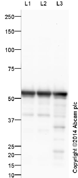 Western blot - Anti-beta III Tubulin antibody - Neuronal Marker (ab18207)All lanes : Anti-beta III Tubulin antibody - Neuronal Marker (ab18207) at 1 µg/mlLane 1 : Brain (Mouse) Tissue LysateLane 2 : Brain (Rat) Tissue LysateLane 3 : Human brain tissue lysate - total protein (ab29466)Lysates/proteins at 10 µg per lane.SecondaryAll lanes : Goat Anti-Rabbit IgG H&L (HRP) (ab97051) at 1/50000 dilutionDeveloped using the ECL technique.Performed under reducing conditions.Predicted band size: 50 kDaObserved band size: 55 kDa why is the actual band size different from the predicted?Exposure time: 30 seconds
This blot was produced using a 4-12% Bis-tris gel under the MOPS buffer system. The gel was run at 200V for 50 minutes before being transferred onto a Nitrocellulose membrane at 30V for 70 minutes. The membrane was then blocked for an hour using 2% Bovine Serum Albumin before being incubated with ab18207 overnight at 4°C. Antibody binding was detected using Goat Anti-Rabbit IgG H&L (HRP) (ab97051) secondary antibody, and visualised using ECL development solution ab133406
 Immunocytochemistry/ Immunofluorescence - Anti-beta III Tubulin antibody - Neuronal Marker (ab18207)
ab18207 staining beta III Tubulin in NGF-differentiated PC12 (Rat adrenal gland pheochromocytoma cell line) cells. The cells were fixed with 100% methanol (5min) and then blocked in 1% BSA/10% normal goat serum/0.3M glycine in 0.1%PBS-Tween for 1h. The cells were then incubated with ab18207 at 5μg/ml and ab7291 at 1µg/ml overnight at +4°C, followed by a further incubation at room temperature for 1h witha Goat Anti-Rabbit IgG H&L (Alexa Fluor® 488) preadsorbed (ab150081) secondary antibodyat 2 μg/ml (shown in green) and Goat Anti-Mouse IgG H&L (Alexa Fluor® 594) (ab150120) secondary antibodyat 2 μg/ml (shown in pseudo color red). Nuclear DNA was labelled in blue with DAPI.Negative controls: 1– Rabbit primary and anti-mouse secondary antibody; 2 – Mouse primary antibody and anti-rabbit secondary antibody. Controls 1 and 2 indicate that there is no unspecific reaction between primary and secondary antibodies used.
 Immunohistochemistry (Formalin/PFA-fixed paraffin-embedded sections) - Anti-beta III Tubulin antibody - Neuronal Marker (ab18207)This image is courtesy of Carl Hobbs, King"s College London, United Kingdomab18207 at 1/2000 staining rat cerebellum tissue sections by Immunohistochemistry (Formalin/PFA-fixed paraffin-embedded sections). The tissue was formaldehyde fixed and a heat mediated antigen retrieval step was performed prior to incubation with the antibody for 16 hours. A biotinylated goat polyclonal antibody was used as the secondary.
See Abreview
 Immunohistochemistry (Formalin/PFA-fixed paraffin-embedded sections) - Anti-beta III Tubulin antibody - Neuronal Marker (ab18207)This image is courtesy of Carl Hobbs, King"s College London, United KingdomImmunohistochemistical staining (Formaldehyde/PFA-fixed paraffin-embedded sections) for Neuron specific beta III Tubulin antibody - Neuronal Marker (ab18207) on Dogfish/Catshark Tissue sections (head: snout region). Antigen retrieval step: Heat mediated. Blocking step:1% BSA for 10 mins at RT. Primary Antibody used at 1/2000 incubated for 2 hours at RT. Secondary Antibody: Biotin labelled goat anti rabbit IgG (1/300).
 Western blot - Anti-beta III Tubulin antibody - Neuronal Marker (ab18207)This image is courtesy of an abreview submitted by Dr Sergi Bayod.All lanes : Anti-beta III Tubulin antibody - Neuronal Marker (ab18207) at 1/1000 dilutionAll lanes : Mouse hippocampus tissue lysateLysates/proteins at 8 µg per lane.SecondaryAll lanes : Goat anti-rabbit IgG (H&L) at 1/5000 dilutionPredicted band size: 50 kDaObserved band size: 55 kDa why is the actual band size different from the predicted?Exposure time: 10 seconds
See Abreview
 Immunocytochemistry/ Immunofluorescence - Anti-beta III Tubulin antibody - Neuronal Marker (ab18207)Sheikh, M.A. et al PLoS One. 2013;8(2):e55826. doi: 10.1371/journal.pone.0055826. Epub 2013 Feb 7 Reproduced under the Creative Commons license http://creativecommons.org/licenses/by/4.0/
Differential expression of Dnmt1, Dnmt3a, and Dnmt3b during RA induced neuronal differentiation of P19 cells
Mouse P19 cells either left untreated (top panel) or RA treated for initial 2 days and further cultured for 4 days without RA (6 days, bottom panel) were immunostained with neuron specific β-III tubulin antibody and nuclei were stained using DAPI.
In order to confirm the neuronal morphology, the cells were stained for neuron specific beta III-tubulin (ab18207). RA induced P19 cells showed immunoreactivity against βIII-tubulin, indicating a neuronal phenotype. In contrast, undifferentiated P19 cells were βIII-tubulin negative.
(After Figure 1A of Sheikh et al)
 Immunohistochemistry (Formalin/PFA-fixed paraffin-embedded sections) - Anti-beta III Tubulin antibody - Neuronal Marker (ab18207)Courtesy of Feng Y et al. Sci Rep. 2017; 7: 44810. doi: 10.1038/srep44810 Reproduced under the Creative Commons license http://creativecommons.org/licenses/by/4.0/.
Immunohistochemical analysis of adult mice ovaries undergone Clarity processing staining tyrosine hydroxlase (TH),Beta III Tubulin (Tuj1) with ab18207, and brain derived neurotrpic factor (BDNF) with ab72439. Positive staing of Tuj1 and BDNF is evident in the theca cells and corpus luteum.
 Immunocytochemistry/ Immunofluorescence - Anti-beta III Tubulin antibody - Neuronal Marker (ab18207)
ab18207 staining beta III tubulin in PC-12 (Rat adrenal gland pheochromocytoma cell line) cells treated with venlafaxine hydrochloride (ab120715), by ICC/IF. Increase in the number and length of neurites (stained with beta III tubulin) correlates with increased concentration of venlafaxine hydrochloride, as described in literature.The NGF treated cells were incubated at 37°C for 6 hour in media containing different concentrations of ab120715 (venlafaxine hydrochloride) in DMSO, fixed with 4% formaldehyde for 10 minutes at room temperature and blocked with PBS containing 10% goat serum, 0.3 M glycine, 1% BSA and 0.1% tween for 2h at room temperature. Staining of the treated cells with ab18207 (1 μg/ml) was performed overnight at 4°C in PBS containing 1% BSA and 0.1% tween. A Goat Anti-Rabbit IgG H&L (DyLight® 488) preadsorbed (ab96899) secondary antibodyat 1/250 dilution was used.
Abcam 位于英国的剑桥科学园,成立于1998年,专门生产和分销研究型抗体。在线目录 (www.abcam.cn) 已有超过 120,000 种抗体和试剂,并不断添加,供应予全球百多个国家。
为使研究员更容易找到蛋白质研究试剂产品,在2011并购了美国的 MitoSciences 公司,加强了免疫分析方面的产品供应;同年也并购了英国的 Ascent Scientific 公司,开展了生化试剂的供应。在2012年并购了美国的 Epitomics 公司,成为一家有领导地位的 RabMAbs® 供应商。
Abcam 的目标是给世界上最好的抗体建立最大的在线目录,为各地科学家提供尖端产品,成为各国科学界的重要伙伴,为所有产品提供技术支持来使客户获得预期的结果,为提供高质量的抗体来指向尽可能多的靶蛋白,尽所能在尽可能多的应用和物种中检测每种抗体。
Abcam 专注于特定的研究领域,包括肿瘤研究、心血管研究、染色质和基因调控研究、免疫学、内参对照、微生物学、神经科学、细胞核信号转导研究、RNAi 研究、干细胞研究、亚细胞标记物等。
![Abcam/Anti-GAPDH antibody [6C5] - Loading Control (ab8245)/1/ab8245](images/Abcam/ab8245-209020-anti-gapdh-antibody-6c5-loading-control-western-blot.jpg)


![Abcam/Recombinant Rabbit monoclonal [M1gG51-4] Anti-Mouse IgG1 H&L (ab125913)/1/ab125913](images/Abcam/ab125913-288990-rabbit-monoclonal-m1gg51-4-anti-mouse-igg1-hampl-elisa.png)
![]()
暂无问答


本网站将在规定时间内给予删除等相关处理。