
Images
 Western blot - Anti-beta Actin antibody (ab8227)All lanes : Anti-beta Actin antibody (ab8227) at 1/1000 dilutionLane 1 : A431 (human epidermoid carcinoma cell line) Whole Cell LysateLane 2 : HEK-293 (human epithelial cell line from embryonic kidney) Whole Cell LysateLane 3 : NIH/3T3 (mouse embryo fibroblast cell line) Whole Cell LysateLane 4 : PC-12 (rat adrenal gland pheochromocytoma cell line) Whole Cell LysateLysates/proteins at 20 µg per lane.SecondaryAll lanes : Goat Anti-Rabbit IgG H&L (Alexa Fluor® 790) (ab175781) at 1/10000 dilutionObserved band size: 42 kDa why is the actual band size different from the predicted?
This blot was produced using a 4-12% Bis-tris gel under the MOPS buffer system. The gel was run at 200V for 50 minutes before being transferred onto anitrocellulose membrane at 30V for 70 minutes. The membrane was then blocked for an hour using 5% Milk before being incubated with ab8227 overnight at 4°C. Antibody binding was detected using Goat Anti-Rabbit IgG H&L (Alexa Fluor® 790) (ab175781) secondary antibody at a 1:10,000 dilution for 1hr at room temperature and then imaged using the Licor Odyssey CLx.
 Immunocytochemistry/ Immunofluorescence - Anti-beta Actin antibody (ab8227)
ab8227 staining beta Actin in SV40LT-SMC (rat aortic smooth muscle cells transfected with SV40). The cells were fixed with 4% formaldehyde (10min), permeabilized with 0.1% Triton X-100 for 5 minutes and then blocked with 1% BSA/10% normal goat serum/0.3M glycine in 0.1%PBS-Tween for 1h. The cells were then incubated overnight at +4°C with ab8227 at 1μg/ml (detected with ab150081, Alexa Fluor® 488 Goat anti-Rabbit, 1μg/ml, shown in green); and ab195889, Mouse monoclonal [DM1A] to alpha Tubulin - Microtubule Marker (Alexa Fluor® 594), at 1/250 dilution (shown in red). Nuclear DNA was labelled with DAPI (shown in blue).Image was taken with a confocal microscope (Leica-Microsystems, TCS SP8).
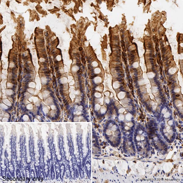
 Immunohistochemistry (Formalin/PFA-fixed paraffin-embedded sections) - Anti-beta Actin antibody (ab8227)
IHC image of ab8227 staining beta Actin in rat small intestine formalin fixed paraffin embedded tissue sections, performed on a Leica Bond. The section was pre-treated using heat mediated antigen retrieval with EDTA (epitope retrieval solution 2) for 20 mins. The section was then incubated with ab8227, 0.2μg/ml, for 15 mins at room temperature and detected using an HRP conjugated compact polymer system. DAB was used as the chromogen. The section was then counterstained with haematoxylin and mounted with DPX. No primary antibody was used in the secondary only control (shown on the inset).For other IHC staining systems (automated and non-automated) customers should optimize variable parameters such as antigen retrieval conditions, primary antibody concentration and antibody incubation times.
 Immunocytochemistry/ Immunofluorescence - Anti-beta Actin antibody (ab8227)
ab8227 staining beta Actin in NIH/3T3 (mouse embryo fibroblast cell line) cells. The cells were fixed with 100% methanol (5min), permeabilized with 0.1% Triton X-100 for 5 minutes and then blocked with 1% BSA/10% normal goat serum/0.3M glycine in 0.1%PBS-Tween for 1h. The cells were then incubated overnight at +4°C with ab8227 at 1μg/ml (shown in green) and ab195889, Mouse monoclonal [DM1A] to alpha Tubulin - Microtubule Marker (Alexa Fluor® 594), at 1/250 dilution (shown in red). Nuclear DNA was labelled with DAPI (shown in blue).Image was taken with a confocal microscope (Leica-Microsystems, TCS SP8).
 Immunohistochemistry (Formalin/PFA-fixed paraffin-embedded sections) - Anti-beta Actin antibody (ab8227)
IHC image of beta actin staining in a section of formalin-fixed paraffin-embeddednormal humancolon*. The section was pre-treated using pressure cooker heat mediated antigen retrieval with sodium citrate buffer (pH6) for 30mins. The section was then incubated with ab8227, 1/1000 dilution, for 15 mins at room temperature. A goat anti-rabbit biotinylated secondary antibody (ab6720, 1/1000 dilution) was used to detect the primary, and visualized using an HRP conjugated ABC system. Streptavidin HRP was used, ab7403 at a 1/10000 dilution. DAB was used as the chromogen (ab103723), diluted 1/100 and incubated for 10min at room temperature. The section was then counterstained with haematoxylin and mounted with DPX.
The inset negative control image is taken from an identical assay without primary antibody.
For other IHC staining systems (automated and non-automated) customers should optimize variable parameters such as antigen retrieval conditions, primary antibody concentration and antibody incubation times.
*Tissue obtained from the Human Research Tissue Bank, supported by the NIHR Cambridge Biomedical Research Centre
 Western blot - Anti-beta Actin antibody (ab8227)All lanes : Anti-beta Actin antibody (ab8227) at 1 µg/mlLane 1 : HeLa (human epithelial cell line from cervix adenocarcinoma) whole cell lysate (blocked with 2% BSA)Lane 2 : NIH/3T3 (mouse embryo fibroblast cell line) whole cell lysate (blocked with 2% BSA)Lane 3 : Rat Liver tissue lysate (blocked with 2% BSA)Lane 4 : HeLa whole cell lysate (blocked with 3% Milk)Lane 5 : NIH/3T3 whole cell lysate (blocked with 3% Milk)Lane 6 : Rat Liver tissue lysate (blocked with 3% Milk)Lysates/proteins at 10 µg per lane.SecondaryAll lanes : Goat Anti-Rabbit IgG H&L (HRP) at 1/50000 dilutionDeveloped using the ECL technique.Performed under reducing conditions.Observed band size: 45 kDa why is the actual band size different from the predicted?Exposure time: 10 seconds
Lanes 1-3: Blocked with 2% BSA
Lanes 4-6: Blocked with 3% Milk
This blot was produced using a 4-12% Bis-tris gel under the MOPS buffer system. The gel was run at 200V for 50 minutes before being transferred onto a Nitrocellulose membrane at 30V for 70 minutes. The membrane was then blocked for an hour using 2% Bovine Serum Albumin or 3% Milk before being incubated with ab8227 overnight at 4°C. Antibody binding was detected using an anti rabbit HRP secondary antibody, and visualised using ECL development solution ab133406
 Western blot - Anti-beta Actin antibody (ab8227)All lanes : Anti-beta Actin antibody (ab8227) at 0.1 µg/mlLane 1 : HeLa (human epithelial cell line from cervix adenocarcinoma) Whole Cell LysateLane 2 : NIH/3T3 (mouse embryonic fibroblast cell line) Whole Cell LysateLane 3 : Liver (Rat) Tissue LysateLysates/proteins at 10 µg per lane.SecondaryAll lanes : Goat Anti-Rabbit IgG H&L (HRP) (ab97051) at 1/50000 dilutionPerformed under reducing conditions.Observed band size: 42 kDa why is the actual band size different from the predicted?Exposure time: 1 minute
This blot was produced using a 4-12% Bis-tris gel under the MOPS buffer system. The gel was run at 200V for 50 minutes before being transferred onto a Nitrocellulose membrane at 30V for 70 minutes. The membrane was then blocked for an hour using 2% Bovine Serum Albumin before being incubated with ab8227 overnight at 4°C. Antibody binding was detected using an anti-rabbit HRP secondary antibody, and visualised using ECL development solution ab133406
 Western blot - Anti-beta Actin antibody (ab8227)All lanes : Anti-beta Actin antibody (ab8227) at 1/1000 dilutionLane 1 : HeLa (human epithelial cell line from cervix adenocarcinoma) nuclear lysateLane 2 : HeLa whole cell lysateLane 3 : A431 (human epidermoid carcinoma cell line) cell lysateLane 4 : Jurkat (human T cell leukemia cell line from peripheral blood) cell lysateLane 5 : HEK-293 (human epithelial cell line from embryonic kidney) cell lysateLane 6 : HeLa nuclear lysate with Human beta Actin peptide (ab13772) at 1 µg/mlLane 7 : HeLa whole cell lysate with Human beta Actin peptide (ab13772) at 1 µg/mlLane 8 : A431 cell lysate with Human beta Actin peptide (ab13772) at 1 µg/mlLane 9 : Jurkat cell lysate with Human beta Actin peptide (ab13772) at 1 µg/mlLane 10 : HEK-293 cell lysate with Human beta Actin peptide (ab13772) at 1 µg/mlLysates/proteins at 20 µg per lane.SecondaryAll lanes : Goat Anti-Rabbit IgG H&L (HRP) (ab6721) at 1/5000 dilutionObserved band size: 41.7 kDa why is the actual band size different from the predicted?Exposure time: 5 seconds
 Western blot - Anti-beta Actin antibody (ab8227)All lanes : Anti-beta Actin antibody (ab8227) at 1/1000 dilutionLane 1 : HeLa (Human epithelial cell line from cervix adenocarcinoma) cellsLane 2 : NIH/3T3 (Mouse embryonic fibroblast cell line) cellsLane 3 : Fish LiverLane 4 : Rabbit LiverLane 5 : MDCK (Canine kidney cell line) cellsLane 6 : EBTr (cow trachea) cellsLane 7 : SL-29 (chicken day 11 embryo) cellsLane 8 : CHO (Chinese hamster ovary cell line) cellsLane 9 : Xenopus laevis embryoLysates/proteins at 20 µg per lane.SecondaryAll lanes : Goat Anti-Rabbit IgG H&L (HRP) (ab6721) at 1/5000 dilutionObserved band size: 41.7 kDa why is the actual band size different from the predicted?Exposure time: 30 seconds
 Immunohistochemistry (Formalin/PFA-fixed paraffin-embedded sections) - Anti-beta Actin antibody (ab8227)
IHC image of beta Actin staining in normal human colon, formalin-fixed and paraffin-embedded tissue*. The section was pre-treated using pressure cooker heat mediated antigen retrieval with sodium citrate buffer (pH6) for 30mins. The section was incubated with ab8227, 3µg/ml overnight at +4°C. A anti-rabbit HRP secondary antibody(Ab97200, 1/200 dilution) was used for 1hr at room temperature. The section was counterstained with haematoxylin and mounted with DPX.
The inset negative control image is taken from an identical assay without primary antibody.
*Tissue obtained from the Human Research Tissue Bank, supported by the NIHR Cambridge Biomedical Research Centre
Abcam 位于英国的剑桥科学园,成立于1998年,专门生产和分销研究型抗体。在线目录 (www.abcam.cn) 已有超过 120,000 种抗体和试剂,并不断添加,供应予全球百多个国家。
为使研究员更容易找到蛋白质研究试剂产品,在2011并购了美国的 MitoSciences 公司,加强了免疫分析方面的产品供应;同年也并购了英国的 Ascent Scientific 公司,开展了生化试剂的供应。在2012年并购了美国的 Epitomics 公司,成为一家有领导地位的 RabMAbs® 供应商。
Abcam 的目标是给世界上最好的抗体建立最大的在线目录,为各地科学家提供尖端产品,成为各国科学界的重要伙伴,为所有产品提供技术支持来使客户获得预期的结果,为提供高质量的抗体来指向尽可能多的靶蛋白,尽所能在尽可能多的应用和物种中检测每种抗体。
Abcam 专注于特定的研究领域,包括肿瘤研究、心血管研究、染色质和基因调控研究、免疫学、内参对照、微生物学、神经科学、细胞核信号转导研究、RNAi 研究、干细胞研究、亚细胞标记物等。


![Abcam/Anti-GAPDH antibody [6C5] - Loading Control (ab8245)/1/ab8245](images/Abcam/ab8245-209020-anti-gapdh-antibody-6c5-loading-control-western-blot.jpg)


![Abcam/Recombinant Rabbit monoclonal [M1gG51-4] Anti-Mouse IgG1 H&L (ab125913)/1/ab125913](images/Abcam/ab125913-288990-rabbit-monoclonal-m1gg51-4-anti-mouse-igg1-hampl-elisa.png)

