


Images
 Western blot - Anti-Histone H3 antibody - Nuclear Marker and ChIP Grade (ab1791)All lanes : Anti-Histone H3 antibody - Nuclear Marker and ChIP Grade (ab1791) at 1/1000 dilutionLane 1 : A431 (Human epithelial carcinoma cell line) Whole Cell LysateLane 2 : Jurkat (Human T cell lymphoblast-like cell line) Whole Cell LysateLane 3 : HEK293 (Human embryonic kidney cell line) Whole Cell LysateLane 4 : A431 (Human epithelial carcinoma cell line) Whole Cell Lysate with Human Histone H3 peptide (ab12149) at 1 µg/mlLane 5 : Jurkat (Human T cell lymphoblast-like cell line) Whole Cell Lysate with Human Histone H3 peptide (ab12149) at 1 µg/mlLane 6 : HEK293 (Human embryonic kidney cell line) Whole Cell Lysate with Human Histone H3 peptide (ab12149) at 1 µg/mlLysates/proteins at 20 µg per lane.SecondaryAll lanes : Goat Anti-Rabbit IgG H&L (HRP) (ab6721) at 1/5000 dilutionDeveloped using the ECL technique.Performed under reducing conditions.Predicted band size: 15 kDaObserved band size: 17 kDa why is the actual band size different from the predicted?Exposure time: 10 seconds
This blot was produced using a 4-12% Bis-tris gel under the MES buffer system. The gel was run at 200V for 35 minutes before being transferred onto a Nitrocellulose membrane at 30V for 70 minutes. The membrane was then blocked for an hour using 2% Bovine Serum Albumin before being incubated with ab1791 overnight at 4°C.
Goat Anti-Rabbit IgG H&L (HRP) (ab6721) secondary antibody was used for detection.
Antibody binding was visualised using ECL development solution ab133406.
 Immunoprecipitation - Anti-Histone H3 antibody - Nuclear Loading Control and ChIP Grade (ab1791)
Histone H3 - ChIP Grade was immunoprecipitated using 0.5mg HeLa (Human epithelial cell line from cervix adenocarcinoma) whole cell extract, 5 µg of Rabbit polyclonal to and 50 µl of protein G magnetic beads (+). No antibody was added to the control (-).
The antibody was incubated under agitation with Protein G beads for 10 minutes, HeLa whole cell extract lysate diluted in RIPA buffer was added to each sample and incubated for a further 10 minutes under agitation.
Proteins were eluted by addition of 40 µl SDS loading buffer and incubated for 10 minutes at 70°C; 10 µl of each sample was separated on a SDS PAGE gel, transferred to a nitrocellulose membrane, blocked with 5% BSA and probed with ab1791.
Secondary Antibody: Mouse anti-rabbit HRP light chain (HRP) (ab99697).
Band: 15kDa; Histone H3 - ChIP Grade
 ChIP - Anti-Histone H3 antibody - Nuclear Loading Control and ChIP Grade (ab1791)
Chromatin from Xenopus laevis oocytes was prepared according to the Abcam X-ChIP protocol.
Oocytes were fixed with formaldehyde for 10 minutes. The ChIP was performed with 25 mg of chromatin, 3 mg of ab7834 (anti-H3, light blue) and 3 µg of ab1791 (anti-H3, dark blue), and 20 ml of Protein A/G sepharose beads. A non-specific antibody was used as a control (yellow).
The immunoprecipitated DNA was quantified by real time PCR (Taqman approach).
 Immunohistochemistry (Formalin/PFA-fixed paraffin-embedded sections) - Anti-Histone H3 antibody - Nuclear Loading Control and ChIP Grade (ab1791)This image is courtesy of an anonymous Abreview
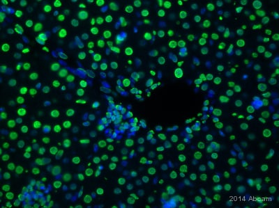
ab1796 staining Histone H3 in mouse liver tissue sections by Immunohistochemistry (IHC-P - paraformaldehyde-fixed, paraffin-embedded sections).
Tissue was fixed with paraformaldehyde, permeabilized with 0.05% Triton X-100 in PBS for30 minutesand blocked with 5% BSA for 1 hour; antigen retrieval was by heat mediation insodium citrate pH 6. Samples were incubated with the primary antibody (1/500 in blocking buffer) for 16 hours at 4°C. An Alexa Fluor® 488-conjugated goat anti-rabbit IgG polyclonal (1/400) was used as the secondary antibody.
See Abreview
 Immunocytochemistry - Anti-Histone H3 antibody - Nuclear Marker and ChIP Grade (ab1791)
ab1791 staining Histone H3 in HeLa cells. The cells were fixed with 100% methanol (5 min), permeabilized with 0.1% PBS-Triton X-100 for 5 minutes and then blocked with 1% BSA/10% normal goat serum/0.3M glycine in 0.1% PBS-Tween for 1h. The cells were then incubated overnight at 4°C with ab1791 at 0.1 µg/mL and ab7291, Mouse monoclonal [DM1A] to alpha Tubulin - Loading Control. Cells were then incubated with ab150081, Goat polyclonal Secondary Antibody to Rabbit IgG - H&L (Alexa Fluor® 488), pre-adsorbed at 1/1000 dilution (shown in green) and ab150120, Goat polyclonal Secondary Antibody to Mouse IgG - H&L (Alexa Fluor® 594), pre-adsorbed at 1/1000 dilution (shown in pseudocolour red). Nuclear DNA was labelled with DAPI (shown in blue).
 Western blot - Anti-Histone H3 antibody - Nuclear Loading Control and ChIP Grade (ab1791)This image is courtesy of an anonymous AbreviewAll lanes : Anti-Histone H3 antibody - Nuclear Marker and ChIP Grade (ab1791) at 1/1000 dilutionLane 1 : Mouse skeletal muscle mitochondrial fractionLane 2 : Mouse skeletal muscle nuclear fractionLysates/proteins at 20 µg per lane.SecondaryAll lanes : HRP-conjugated goat anti-rabbit IgG at 1/4000 dilutionDeveloped using the ECL technique.Performed under reducing conditions.Predicted band size: 15 kDaObserved band size: 17 kDa why is the actual band size different from the predicted?Exposure time: 7 minutes
Blocked with 3% milk for 1 hour at 25°C.
Incubated with the primary antibody for 16 hours at 4°C in 3% milk in TBS-tween.
See Abreview
 Immunocytochemistry - Anti-Histone H3 antibody - Nuclear Marker and ChIP Grade (ab1791)This image is courtesy of an anonymous Abreview
ab1791 staining Histone H3 in HeLa (Human epithelial cell line from cervix adenocarcinoma) by ICC/IF (Immunocytochemistry/immunofluorescence).
Cells were fixed with methanol and blocked with 0.2% fish scale gelatin for 1 hour at 25°C. Samples were incubated with the primary antibody (1/300 in PBS + 0.2% gelatin) for 20 minutes at 25°C. An Alexa Fluor® 488-conjugated donkey anti-rabbit IgG polyclonal (1/500) was used as the secondary antibody.
Green - Histone H3.Blue - DAPI.Red - Tubulin.
See Abreview
 Western blot - Anti-Histone H3 antibody - Nuclear Loading Control and ChIP Grade (ab1791)All lanes : Anti-Histone H3 antibody - Nuclear Marker and ChIP Grade (ab1791) at 1 µg/mlLane 1 : HeLa (Human epithelial carcinoma cell line) Whole Cell LysateLane 2 : NIH 3T3 whole cell lysate (ab7179)Lane 3 : Drosophila embryo nuclear extract (from melanogaster embryos 0-12Hr)Lane 4 : S.cerevisiae (Y190) Whole Cell Lysate Lane 5 : S.pombe Whole Cell Lysate Lysates/proteins at 10 µg per lane.SecondaryAll lanes : Goat polyclonal to Rabbit IgG - H&L - Pre-Adsorbed (HRP) at 1/3000 dilutionPerformed under reducing conditions.Predicted band size: 15 kDaObserved band size: 17 kDa why is the actual band size different from the predicted?ab1791 is tested in western blot on a range of species. We recommend loading higher amounts of protein (20-30ug) to increase the signal in yeast lysates
 Western blot - Anti-Histone H3 antibody - Nuclear Loading Control and ChIP Grade (ab1791)This image is courtesy of John E. Mueller and J. Ruth German (Mary Bryk lab)
Rabbit polyclonal to Histone H3 (ab1791) at 1/5000 on S. cerevisiae whole cell lysate (40 ug per lane). Protein resolved on 15% SDS-PAGE gel. After transfer to PVDF membrane, blots were blocked in 1X PBS, 0.1% Tween-20, and 5% milk. ab1791 was diluted in 5 ml blocking buffer at 1/5000. Blots plus primary antibodies were either incubated overnight at 4°C or at RT for 2 hours. Blots were washed 6X for 10 minutes each in PBS with 0.1% Tween-20 before addition of secondary antibodies. Secondary antibodies were diluted 1/2,000 in blocking buffer and incubated with blots for 2 hours at RT. Secondary blots were washed 4X for 10 minutes each in PBS with 0.1% Tween-20 and 2X for 10 minutes each in PBS.
 ChIP - Anti-Histone H3 antibody - Nuclear Loading Control and ChIP Grade (ab1791)
Chromatin was prepared from HeLa (Human epithelial cell line from cervix adenocarcinoma) cells according to the Abcam X-ChIP protocol.
Cells were fixed with formaldehyde for 10 minutes. The ChIP was performed with 25 µg of chromatin, 2 µg of ab1791 (blue), and 20 µl of Protein A/G sepharose beads. No antibody was added to the beads control (yellow).
The immunoprecipitated DNA was quantified by real time PCR (Taqman approach for active and inactive loci, Sybr green approach for heterochromatic loci). Primers and probes are located in the first kb of the transcribed region.
 ChIP - Anti-Histone H3 antibody - Nuclear Loading Control and ChIP Grade (ab1791)
The ChIP was performed with chromatin from mouse gut cell lysate and ab1791at 1/250 dilution.
Negative control: Noantibodywas used (right bar).
The immunoprecipitated DNA was quantified by real time PCR.
See Abreview
 Immunohistochemistry (Formalin/PFA-fixed paraffin-embedded sections) - Anti-Histone H3 antibody - Nuclear Loading Control and ChIP Grade (ab1791)
Paraffin-embedded rat brain tissue stained for Histone H3 using ab1791 at 1/8000 dilution in immunohistochemical analysis.
See Abreview
 Immunohistochemistry (Formalin/PFA-fixed paraffin-embedded sections) - Anti-Histone H3 antibody - Nuclear Loading Control and ChIP Grade (ab1791)This image is courtesy of an anonymous Abreview
ab1791 staining Histone H3 in human infantile fibromatosis tissue sections by Immunohistochemistry (IHC-P - paraformaldehyde-fixed, paraffin-embedded sections).
Tissue was fixed with formaldehyde and blocked with 1% FBS/BSA for 3 hours at room temperature; antigen retrieval was by heat mediation in Tris pH 9. Samples were incubated with primary antibody (1/100 in TBS + 1% BSA + 1% FBS) for 16 hours. An un-dilutedHRP-conjugated goat anti-rabbit IgG polyclonal was used as the secondary antibody.
See Abreview
 Immunohistochemistry (Formalin/PFA-fixed paraffin-embedded sections) - Anti-Histone H3 antibody - Nuclear Loading Control and ChIP Grade (ab1791)
ab1791 staining Histone H3 (red)in rat brain tissue sections by Immunohistochemistry (IHC-P - paraformaldehyde-fixed, paraffin-embedded sections).
Tissue was fixed with formaldehyde, permeabilized with 0.1% TBS-TritonXand blocked with 10% serum for 1 hour at 25°C; antigen retrieval was by heat mediation in a citrate buffer. Samples were incubated with the primary antibody (1/500 in 10% normal goat serum) for 24 hours at 24°C. An Alexa Fluor® 594-conjugated goat anti-rabbit IgG polyclonal (1/500) was used as the secondary antibody.
Green - Nucleus staining.Red - Histone H3 staining.
Abcam 位于英国的剑桥科学园,成立于1998年,专门生产和分销研究型抗体。在线目录 (www.abcam.cn) 已有超过 120,000 种抗体和试剂,并不断添加,供应予全球百多个国家。
为使研究员更容易找到蛋白质研究试剂产品,在2011并购了美国的 MitoSciences 公司,加强了免疫分析方面的产品供应;同年也并购了英国的 Ascent Scientific 公司,开展了生化试剂的供应。在2012年并购了美国的 Epitomics 公司,成为一家有领导地位的 RabMAbs® 供应商。
Abcam 的目标是给世界上最好的抗体建立最大的在线目录,为各地科学家提供尖端产品,成为各国科学界的重要伙伴,为所有产品提供技术支持来使客户获得预期的结果,为提供高质量的抗体来指向尽可能多的靶蛋白,尽所能在尽可能多的应用和物种中检测每种抗体。
Abcam 专注于特定的研究领域,包括肿瘤研究、心血管研究、染色质和基因调控研究、免疫学、内参对照、微生物学、神经科学、细胞核信号转导研究、RNAi 研究、干细胞研究、亚细胞标记物等。
![Abcam/Anti-GAPDH antibody [6C5] - Loading Control (ab8245)/1/ab8245](images/Abcam/ab8245-209020-anti-gapdh-antibody-6c5-loading-control-western-blot.jpg)


![Abcam/Recombinant Rabbit monoclonal [M1gG51-4] Anti-Mouse IgG1 H&L (ab125913)/1/ab125913](images/Abcam/ab125913-288990-rabbit-monoclonal-m1gg51-4-anti-mouse-igg1-hampl-elisa.png)
![]()
暂无问答


本网站将在规定时间内给予删除等相关处理。